Shoulder Joint Anatomy Diagram Easy - Types Of Synovial Joints Biology For Majors Ii : Robin smithuis and henk jan van der woude.
byAdmin•
0
Shoulder Joint Anatomy Diagram Easy - Types Of Synovial Joints Biology For Majors Ii : Robin smithuis and henk jan van der woude.. Shoulder surgery recovery shoulder anatomy joint replacement shoulder injuries knee surgery rotator cuff. Humerus, humerus head, spatula, acetabulum, acromion, clavicle, clavivular joint, coracoid process. Use the mouse scroll wheel to move the images up and down alternatively use the tiny arrows (>>) on both side of the image to move the images. Webmd's shoulder anatomy page provides an image of the parts of the shoulder and describes its function, shoulder problems, and more. The 3b scientific® anatomy video shoulder joint vividly describes the functional and topographical anatomy of the shoulder.
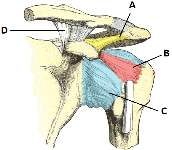
Posted on december 13, 2018december 12, 2018. Equally extensive are the muscles affecting the shoulder movement, including: Three bones come together at the shoulder joint. The shoulder joint is vulnerable to dislocations from sudden jerks of the arm, especially in children before strong muscles have developed. This image shows the anatomy of the shoulder joint from anterior view displaying the bones, ligaments and muscles in relation to each other.
The Shoulder Joint Structure Movement Teachmeanatomy from teachmeanatomy.info The shoulder joint is vulnerable to dislocations from sudden jerks of the arm, especially in children before strong muscles have developed. Various types of injuries and degenerative conditions can cause the shoulder to become painful. Relevant anatomy, mechanism of injury and pathophysiology the carpometacarpal joint is between the base. The shoulder joint is formed where the humerus (upper arm bone) fits into the scapula. In human anatomy, the shoulder joint comprises the part of the body where the humerus attaches to the scapula.1 there are two kinds of cartilage in the joint. It is named after silvio rolando, an italian surgeon who described it first. Shoulder joint is the most mobile joint of the human body. • during abduction of the shoulder joint, the supraspinatus tendon is exposed to friction against the acromion.
This incongruent bony anatomy allows for the wide range of movement available at the shoulder joint but is also the reason for the lack of joint stability.
How to draw heart diagram in exams ? Due to the tension by the anterior band of the inferior ghl labral teras will be easier to detect. It is named after silvio rolando, an italian surgeon who described it first. Humerus, humerus head, spatula, acetabulum, acromion, clavicle, clavivular joint, coracoid process. Start studying shoulder joint anatomy. Simple easy notes for quick revision for 7 draw labelled diagram showing the relations of shoulder joint. Three bones come together at the shoulder joint. The shoulder joint is the most mobile joint in the human body and responsible for movements of arm and scapula. The first type is the white cartilage on the ends of the bones (called articular cartilage) which allows the bones to glide and move on each other. 1 this mobility provides the upper extremity with tremendous range of motion such as adduction, abduction, flexion, extension, internal rotation, external rotation, and 360° circumduction in. Simply put, the shoulder, or shoulder joint, is the connection of the upper arm and the thorax. The shoulder joint is vulnerable to dislocations from sudden jerks of the arm, especially in children before strong muscles have developed. Shoulder joint of human body anatomy infographic diagram with all parts including bones ligaments muscles bursa cavity capsule cartilage membrane for medical science education and health care.
Please support and like, comment,share the video to your. Equally extensive are the muscles affecting the shoulder movement, including: The shoulder joint is formed where the humerus (upper arm bone) fits into the scapula. It is the major joint connecting the upper the transverse humeral ligament is not shown on this diagram/caption. Simply put, the shoulder, or shoulder joint, is the connection of the upper arm and the thorax.
Shoulder Joint Glenohumeral Joint 3d Anatomy Tutorial Youtube from i.ytimg.com Shoulder surgery recovery shoulder anatomy joint replacement shoulder injuries knee surgery rotator cuff. The shoulder joint (glenohumeral joint) is a ball and socket joint between the scapula and the humerus. Erythrocyte sedimentation rate (esr) by shabab ali 21093 views. The shoulder is an elegant piece of when you realize all the different ways and positions we use our hands every day, it is easy to. Various types of injuries and degenerative conditions can cause the shoulder to become painful. Relevant anatomy, mechanism of injury and pathophysiology the carpometacarpal joint is between the base. Human kidney anatomy_easy steps to draw. The 3b scientific® anatomy video shoulder joint vividly describes the functional and topographical anatomy of the shoulder.
Learn vocabulary, terms and more with flashcards, games and other study tools. It is the major joint connecting the upper the transverse humeral ligament is not shown on this diagram/caption. Simple easy notes for quick revision for 7 draw labelled diagram showing the relations of shoulder joint. Due to the tension by the anterior band of the inferior ghl labral teras will be easier to detect. Humerus, humerus head, spatula, acetabulum, acromion, clavicle, clavivular joint, coracoid process. In human anatomy, the shoulder joint comprises the part of the body where the humerus attaches to the scapula.1 there are two kinds of cartilage in the joint. The glenohumeral joint (shoulder joint) is a synovial ball and socket articulation anatomy ▶ upper limb ▶ joints ▶ shoulder joint (glenohumeral joint). 1 this mobility provides the upper extremity with tremendous range of motion such as adduction, abduction, flexion, extension, internal rotation, external rotation, and 360° circumduction in. The first type is the white cartilage on the ends of the bones (called articular cartilage) which allows the bones to glide and move on each other. 8 name the arteries and the nerves that supply shoulder joint. Shoulder joint of human body anatomy infographic diagram with all parts including bones ligaments muscles bursa cavity capsule cartilage membrane for medical science education and health care. Coracoclavicular ligament 3 shoulder joint anatomy. Click now and learn everything about its anatomy and function at kenhub!
Coracoclavicular ligament 3 shoulder joint anatomy. The shoulder joint is the connection between the chest and the upper extremity. Equally extensive are the muscles affecting the shoulder movement, including: 1 this mobility provides the upper extremity with tremendous range of motion such as adduction, abduction, flexion, extension, internal rotation, external rotation, and 360° circumduction in. As a ball and socket synovial joint, there is a wide range of.
9 6 Anatomy Of Selected Synovial Joints Anatomy Physiology from open.oregonstate.education This diagram here just shows the joint capsule itself. Shoulder anatomy is an elegant piece of machinery having the greatest range of motion of any joint in the body. Please support and like, comment,share the video to your. Shoulder joint anatomy,easiest explanation and revise anatomy. All about the shoulder muscles. Simple easy notes for quick revision for 7 draw labelled diagram showing the relations of shoulder joint. Three bones come together at the shoulder joint. Home > blog > anatomy > shoulder anatomy:
As a ball and socket synovial joint, there is a wide range of.
Various types of injuries and degenerative conditions can cause the shoulder to become painful. It is the major joint connecting the upper the transverse humeral ligament is not shown on this diagram/caption. The shoulder joint is the most mobile joint in the human body and responsible for movements of arm and scapula. The shoulder is one of the largest and most complex joints in the body. The 3b scientific® anatomy video shoulder joint vividly describes the functional and topographical anatomy of the shoulder. Related online courses on physioplus. In human anatomy, the shoulder joint comprises the part of the body where the humerus attaches to the scapula.1 there are two kinds of cartilage in the joint. Robin smithuis and henk jan van der woude. Describe the structure of the shoulder should begin with bone parts that include: The shoulder joint by quan fu gan 67157 views. The shoulder joint is the connection between the chest and the upper extremity. Coracoclavicular ligament 3 shoulder joint anatomy. Human kidney anatomy_easy steps to draw.
The shoulder joint by quan fu gan 67157 views shoulder anatomy diagram. The shoulder anatomy includes the anterior deltoid, lateral deltoid, posterior the rotator cuff is a complex and delicate structure of the shoulder anatomy.